Introduction
Dermal fillers have become a popular choice for non-surgical cosmetic enhancement in the UK, offering a way to restore facial volume, smooth out wrinkles, and enhance features without the need for invasive procedures. While generally safe and well-tolerated, dermal fillers can sometimes lead to complications. Among these, late-onset nodules are a concern that can arise weeks, months, or even years after the initial treatment. Understanding the causes, diagnosis, and management of these nodules is crucial for both practitioners and patients to ensure the best possible outcomes.
What Are Late-Onset Nodules?
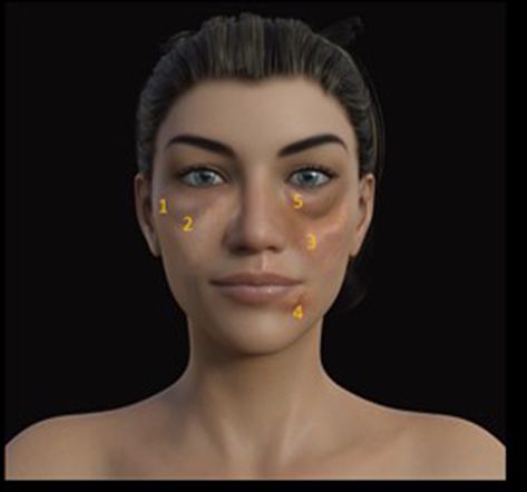
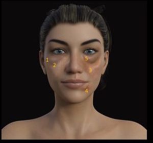
Late-onset nodules refer to the development of lumps or bumps at the site of dermal filler injection that appear after a significant delay, typically weeks to years following the procedure. These nodules can vary in size, consistency, and level of discomfort, ranging from small, painless bumps to larger, painful, and inflamed lesions. The occurrence of these nodules, though relatively rare, can be distressing for patients and challenging for practitioners to treat.
Causes of Late-Onset Nodules
The development of late-onset nodules after dermal filler treatment can be attributed to several factors, including inflammatory reactions, infections, and the type of filler used.
- Immune-Mediated Inflammatory Reactions:
- One of the most common causes of late-onset nodules is a delayed immune-mediated inflammatory response. This can occur when the body’s immune system reacts to the filler material as a foreign substance, even long after the initial treatment. This reaction can lead to granuloma formation, where clusters of immune cells surround the filler material, creating a palpable nodule.
- Biofilm Formation:
- Biofilm refers to a thin layer of bacteria that can form on the surface of the filler material. These bacteria are often protected from the body’s immune system by a slimy matrix, making them resistant to antibiotics. Biofilms can remain dormant for extended periods and may be triggered into activity by factors such as illness, injury, or additional cosmetic procedures, leading to the formation of late-onset nodules.
- Type of Filler:
- The type of dermal filler used can influence the likelihood of developing late-onset nodules. For instance, non-biodegradable fillers, such as polymethylmethacrylate (PMMA) or silicone, are more likely to cause long-term complications compared to biodegradable fillers like hyaluronic acid. Even with biodegradable fillers, certain formulations or impurities can increase the risk of delayed reactions.
- Infection:
- Though less common, infections can also cause late-onset nodules. This can occur if bacteria are introduced at the time of injection or if they spread to the filler site from another part of the body. Infections may remain asymptomatic for a period before manifesting as nodules accompanied by redness, tenderness, or abscess formation.
- Procedural Factors:
- The technique used during filler injection can also play a role in nodule formation. Improper injection techniques, such as injecting too superficially, uneven distribution of the filler, or using unsterile equipment, can increase the risk of complications, including the development of nodules.
Diagnosis of Late-Onset Nodules
Accurate diagnosis of late-onset nodules is essential for determining the appropriate management approach. Diagnosis typically involves a combination of patient history, clinical examination, and sometimes imaging or laboratory tests.
- Patient History: A detailed patient history is the first step in diagnosing late-onset nodules. Practitioners should inquire about the timeline of nodule development, any preceding events (such as illness or trauma), and the type of filler used. Information about previous treatments, including any history of similar reactions, is also valuable.
- Clinical Examination: A physical examination of the affected area is crucial. The practitioner should assess the size, consistency, and tenderness of the nodules, as well as any associated signs of infection, such as redness or warmth. The location of the nodules relative to the original injection sites can also provide clues about their cause.
- Imaging: In some cases, imaging studies such as ultrasound or MRI may be necessary to evaluate the extent of the nodule and to distinguish between different types of tissue reactions. Ultrasound can be particularly useful in identifying the presence of granulomas or abscesses.
- Laboratory Tests: If an infection is suspected, laboratory tests such as blood cultures or swabs of any discharge may be conducted to identify the causative organism. This information is crucial for guiding antibiotic therapy.
- Histopathological Examination: In rare cases, a biopsy of the nodule may be required for histopathological examination. This can help differentiate between granulomatous reactions, infections, and other possible causes of the nodules.
Management of Late-Onset Nodules
The management of late-onset nodules depends on the underlying cause and the severity of the symptoms. Treatment options can range from conservative approaches to more invasive procedures.
- Conservative Management:
- In cases where the nodules are small, asymptomatic, and non-inflamed, conservative management may be appropriate. This can include observation, as some nodules may resolve spontaneously over time. Patients should be advised to monitor the nodules for any changes in size, pain, or appearance.
- Medical Therapy:
- Corticosteroids: For nodules caused by inflammatory reactions, intralesional corticosteroid injections can help reduce inflammation and shrink the nodules. Oral corticosteroids may also be prescribed in more severe cases.
- Antibiotics: If an infection is suspected or confirmed, a course of antibiotics is necessary. The choice of antibiotic should be guided by culture results where possible, and treatment duration may vary depending on the severity of the infection.
- Hyaluronidase: For nodules resulting from hyaluronic acid fillers, hyaluronidase injections can be used to dissolve the filler. This enzyme breaks down hyaluronic acid, which can help resolve the nodule more quickly. However, this approach is not suitable for non-hyaluronic acid fillers.
- Minimally Invasive Procedures:
- Aspiration: If the nodule is fluid-filled or an abscess has formed, aspiration with a fine needle can help to drain the contents and reduce the size of the nodule. This procedure is often guided by ultrasound to ensure accuracy.
- Incision and Drainage: In cases where nodules are large, painful, or do not respond to aspiration, a small incision may be made to drain the contents. This is typically performed under local anaesthesia.
- Surgical Excision:
- Surgical excision may be considered for persistent nodules that do not respond to other treatments or for nodules caused by non-biodegradable fillers. This approach is more invasive and may result in scarring, so it is usually reserved for cases where other treatments have failed.
- Patient Education and Follow-Up:
- Educating patients about the potential risks of dermal fillers, including the possibility of late-onset nodules, is essential. Patients should be informed about the signs and symptoms of complications and the importance of seeking prompt medical attention if they notice any changes.
- Regular follow-up appointments are recommended for patients who have experienced late-onset nodules to monitor their progress and adjust treatment as needed.
Conclusion
Late-onset nodules are a rare but significant complication of dermal filler treatments. While the exact cause of these nodules can vary, a thorough understanding of the potential risks, early diagnosis, and appropriate management strategies are crucial for ensuring patient safety and satisfaction. Clients should be so careful to ensure that they choose an aesthetic practitioner that is able to identify and treat this complication if it arises. Please see my blog which addresses finding the right practitioner. Practitioners should be vigilant in monitoring for signs of complications and be prepared to intervene quickly and effectively when necessary. By doing so, the risks associated with dermal fillers can be minimized, allowing patients to enjoy the aesthetic benefits of these treatments with greater confidence.